Contents
Scroll to:
 Karen M. Nikogosyan,
Marina P. Sutunkova,
Ilzira A. Minigalieva,
Natalia A. Gertan,
Ekaterina M. Petrunina,
Renata R. Sakhautdinova
Karen M. Nikogosyan,
Marina P. Sutunkova,
Ilzira A. Minigalieva,
Natalia A. Gertan,
Ekaterina M. Petrunina,
Renata R. Sakhautdinova https://doi.org/10.47470/0869-7922-2026-34-2-124-131
EDN: xsvivv
Scroll to:
Introduction. Current challenges in toxicology include the development and modernization of biological prevention strategies to reduce the risks of long-term toxic effects of lead.
The aim of this study was to determine changes in certain biochemical and histomorphological parameters of the reproductive system of male rats following subchronic exposure to lead acetate and to establish efficacy of a bioprophylactic complex (hereinafter referred to as BPC) specially developed to reduce the severity of toxic effects.
Material and methods. The experiment was conducted on albino outbred male rats aged from 4 to 6 months. The administration was carried out orally, daily for 45 days. The dose of lead acetate was 819 mg/L. Some of the animals received a vitamin and mineral complex, the composition of which was selected based on the mechanisms of lead toxicity. The effectiveness of BPC and the toxic effects of lead acetate were evaluated 21 days after the end of the exposure.
Results. According to the obtained data, lead exposure resulted in a significant decrease in the lumen of the seminiferous tubules and an increase in the proportion of Sertoli cells with degenerative and dystrophic changes. A significantly higher number of abnormal spermatozoa in testicular smears was detected in rats treated with lead acetate compared with control. An increase in the activity of the enzyme 3β-hydroxysteroid dehydrogenase was observed in exposed rats compared to controls.
Although this study did not measure sex hormone levels in the blood of laboratory animals, we believe that the observed increase in HSD3b1 is more likely a consequence of compensatory responses to the destruction and/or dysfunction of cells responsible for androgen synthesis than a direct consequence of lead toxicity.
Limitations. We did not assess sex hormone levels or the activity of the HSD3b2 isoform of the 3bHSD enzyme.
Conclusion. Male rats exposed to lead acetate showed a slight increase in the activity of 3β-hydroxysteroid dehydrogenase, an enzyme that plays an important role in regulating sex hormone synthesis and delta-ALA metabolism. The observed histomorphometric changes included a decrease in the lumen of the seminiferous tubules, an increase in the number of abnormal Sertoli cells and degenerated spermatozoa. Our findings prove the protective potential of biological prevention strategies but highlight the importance of further profound studies of the mechanisms of lead toxicity on the male reproductive system and the effects of BPC.
Compliance with ethical standards. The study was approved by the local Bioethics Committee of the Yekaterinburg Medical Research Center for Prophylaxis and Health Protection of Industrial Workers of Rospotrebnadzor (protocol 1A of February 3, 2025) and conducted in compliance with the European Convention for the Protection of Vertebrate Animals used for Experimental and other Scientific Purposes (ETS No. 123) and Directive 2010/63/EC of the European Parliament and of the Council of September 22, 2010 on the protection of animals used for scientific purposes.
Authors’ contribution:
Nikogosyan K.M. – data collection and processing, writing, editing of the article;
Sutunkova M.P. – study design and editing;
Minigalieva I.A. – study conception and editing;
Gertan N.A. – data collection and processing;
Petrunina E.M., Sakhautdinova R.R. – data collection.
All co-authors approved the final version of the article and are responsible for the integrity of all its parts.
Conflict of interest. The authors declare the absence of obvious and potential conflicts of interest in connection with the publication of this article.
Funding. The study had no sponsorship.
Received: March 11, 2026 / Accepted: March 27, 2026 / Published: April 30, 2026
Nikogosyan K.M., Sutunkova M.P., Minigalieva I.A., Gertan N.A., Petrunina E.M., Sakhautdinova R.R. Changes in some parameters of the reproductive system of male rats following lead exposure and the effect of bioprophylaxis. Toxicological Review. 2026;34(2):124-131. https://doi.org/10.47470/0869-7922-2026-34-2-124-131. EDN: xsvivv
Introduction
Among the reasons for Russia’s low demographic potential, exposure to harmful chemical factors of various origins occupies a significant place. The population of regions with developed production is traditionally exposed to a high complex chemical load in the habitat and working area¹. In this case, lead acts as one of the most widespread industrial pollutants that contaminate the environment, causing health risks for both humans and their offspring. Many studies have been conducted on the toxic effects of inorganic lead compounds, but the long-term effects as a serious threat to the health of future generations have not been sufficiently studied.
A key role in combating high chemical exposure is played by the strategy of biological prevention on the population, which increases the body’s resistance, adaptation, and elimination capabilities. The developed bioprophylactic complexes (hereinafter referred to as BPC), which include harmless substances with protective properties, have demonstrated their effectiveness in previous experiments implemented on the basis of the Federal State Scientific Institution ‘Yekaterinburg Medical Research Center for Prevention and Health Promotion of Industrial Workers’ of Rospotrebnadzor [1–3]. Current challenges in toxicology include the development and modernization of biological prevention strategies to reduce the risks of long-term toxic effects of lead.
The aim of this study was to determine changes in certain biochemical and histomorphological parameters of the reproductive system of male rats following subchronic exposure to lead acetate and to establish efficacy of a bioprophylactic complex (hereinafter referred to as BPC) specially developed to reduce the severity of these toxic effects
Material and methods
The study was approved by the Local Bioethics Committee of the Federal State Scientific Institution ‘Yekaterinburg Medical Research Center for Prevention and Health Promotion of Industrial Workers’ of Rospotrebnadzor (protocol LEC No. 1A dated February 3, 2025). The experiment was conducted on albino outbred male rats aged from 4 to 6 months (Andreevka branch of the Federal State Scientific Institution ‘Scientific Center for Biomedical Technologies of the Federal Medical and Biological Agency of Russia’, 141551, Moscow Region, Solnechnogorsk, Andreevka settlement, bldg. 49. Certificate No. 181164, issue date: 09.12.2024) Before the experiment, the rats were kept in quarantine, after which they were allowed to enter the vivarium, which was equipped as per GOST 33216–2014². The rats received specialized diet as per GOST R 50258–92³ and purified drinking water. The temperature, humidity, and air flow were carefully controlled to ensure comfort and safety for the laboratory animals during the experiment. A day-night cycle was artificially created in the animal housing areas, simulating sunset and sunrise. Daily veterinary inspections were conducted throughout the experiment.
Four groups, each consisting of eight male rats, were formed for the experiment.
For the composition of the bioprophylactic complex and dosages for laboratory animals, see Table.
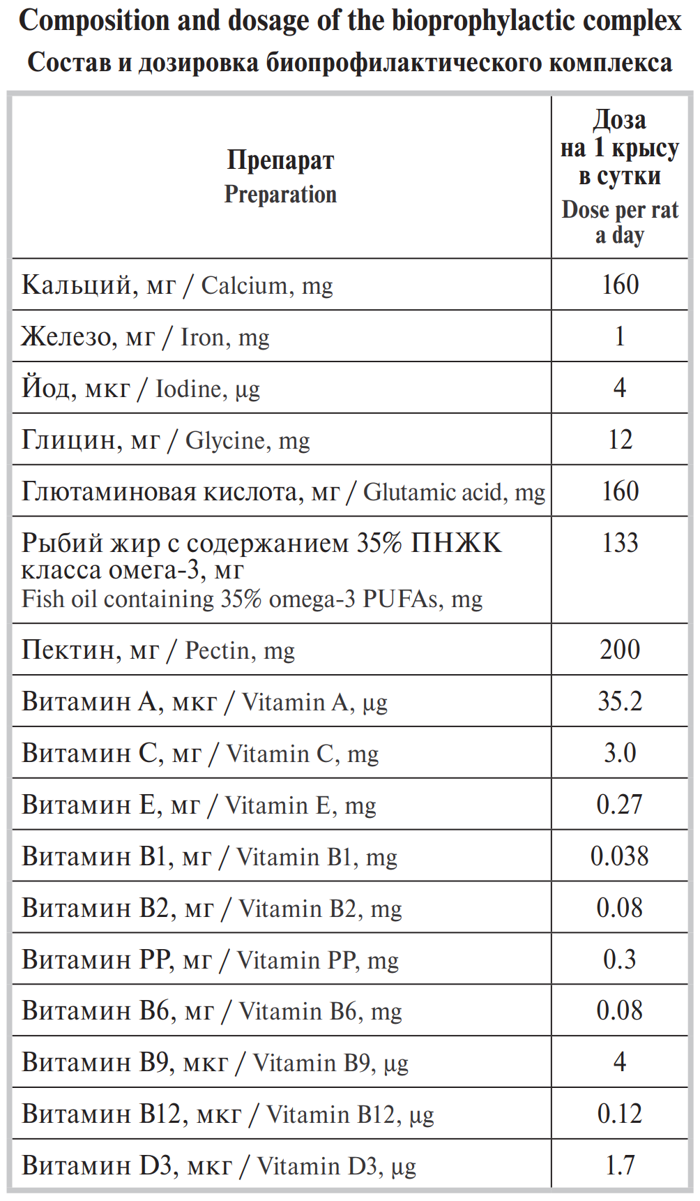
Collected whole blood from the rats twenty-one days after the end of exposure for subsequent assessment of the content of the HSD3b1 isoform of the enzyme 3β-hydroxysteroid dehydrogenase (hereinafter referred to as 3bHSD) in the serum, as well as the testes for morphometric analysis of histological specimens (hematoxylin and eosin staining) and cytomorphological analysis of smears (Leishman staining). Determined HSD3b1 isoform content using an enzyme-linked immunosorbent assay according to the kit instructions. Evaluated during the histomorphological analysis changes in seminiferous tubule lumen, sperm head angle, and counted abnormal sperm forms and Sertoli cells with degenerative-dystrophic changes.
Performed statistical analysis using the Student’s t-test and the Mann-Whitney U-test. Evaluated data normality using the Shapiro-Wilk test. The differences were considered statistically significant with a probability of at least 95% (p < 0.05).
Results
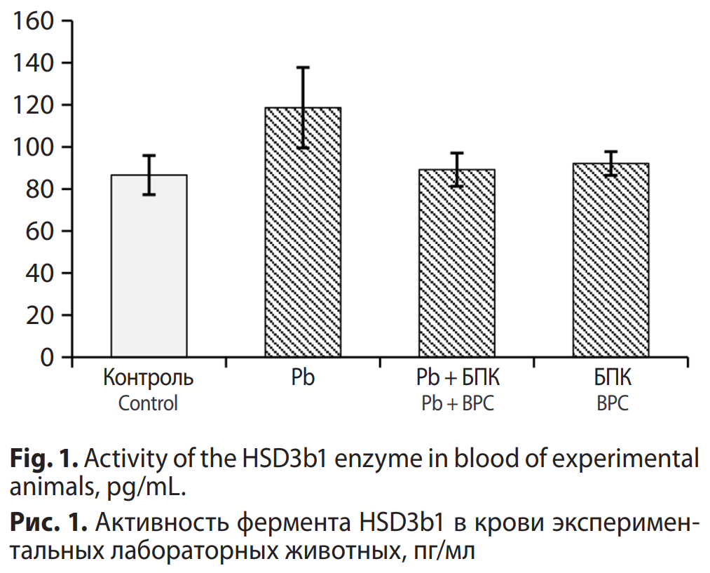
The study revealed that content of the HSD3b1 isoform of the 3bHSD enzyme exhibited a 1.5-fold increase in the Pb rat group compared to the Control, though these differences did not attain statistical significance. The rats of the Pb + BPC and BPC groups demonstrated values of this indicator equivalent to those of the Control (Fig. 1).
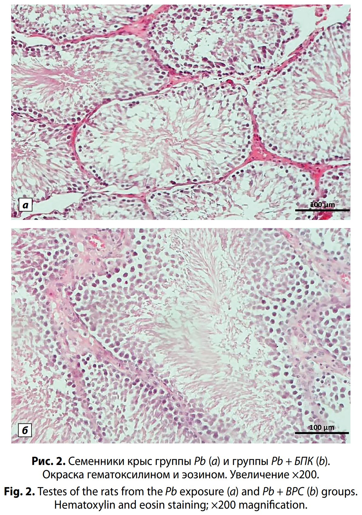
A comparison of the histological pattern of the testes from the Control and the BPC group demonstrated no significant differences. Pathological morphological changes were not observed in either group. Microscopic analysis of histological sections showed that the testicular fibrous membrane consists of a thin layer of connective tissue. The organ’s parenchyma is composed of convoluted seminiferous tubules with rounded or ellipsoid shapes, depending on the orientation of the passing section (either longitudinal or transverse). The outer sheath of the tubules is a very thin layer of annular connective tissue, with a smooth layer of myoid cells along the inner contour, including an inner myoid layer directly adjacent to the basement membrane and a layer of fibroblast-like cells. The spermatogenic epithelium layer is evenly distributed. Different stages of spermatogenesis were observed depending on the level of the sections. Nevertheless, all layers were preserved in almost all tubules: spermatozoa and late forms of spermatids were present in the greatest number, and Leydig cells were detected in the periphery of blood vessels in the amount of 3–5 cells per field of view.
The histological analysis of the Pb group showed a slight reduction in the number of spermatozoa and spermatocytes compared to the Control. This suggested a marginally lower level of spermatogenesis. In contrast to the Controlи BPC groups, fewer Leydig cells were observed in the interstitium: 2–3 per field of view. At the same time, in thePb + BPC group, the changes were less pronounced than in the Pb group. The histological analysis showed that the number of spermatozoa and spermatocytes was slightly lower than in the Control, but higher than in the experimental group. A higher number of Leydig cells was also observed compared to the Pb group: 3–5 per field of view (Fig. 2, see on the insert).
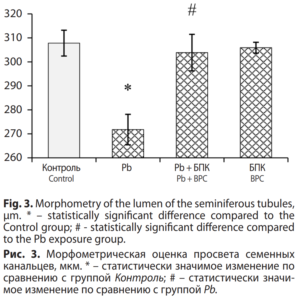
According to the obtained data, lead exposure resulted in a significant decrease in the lumen of the seminiferous tubules in the Pb group. In the Pb + BPC group, changes were minor and consistent with those in the Control and BPC group (Fig. 3).
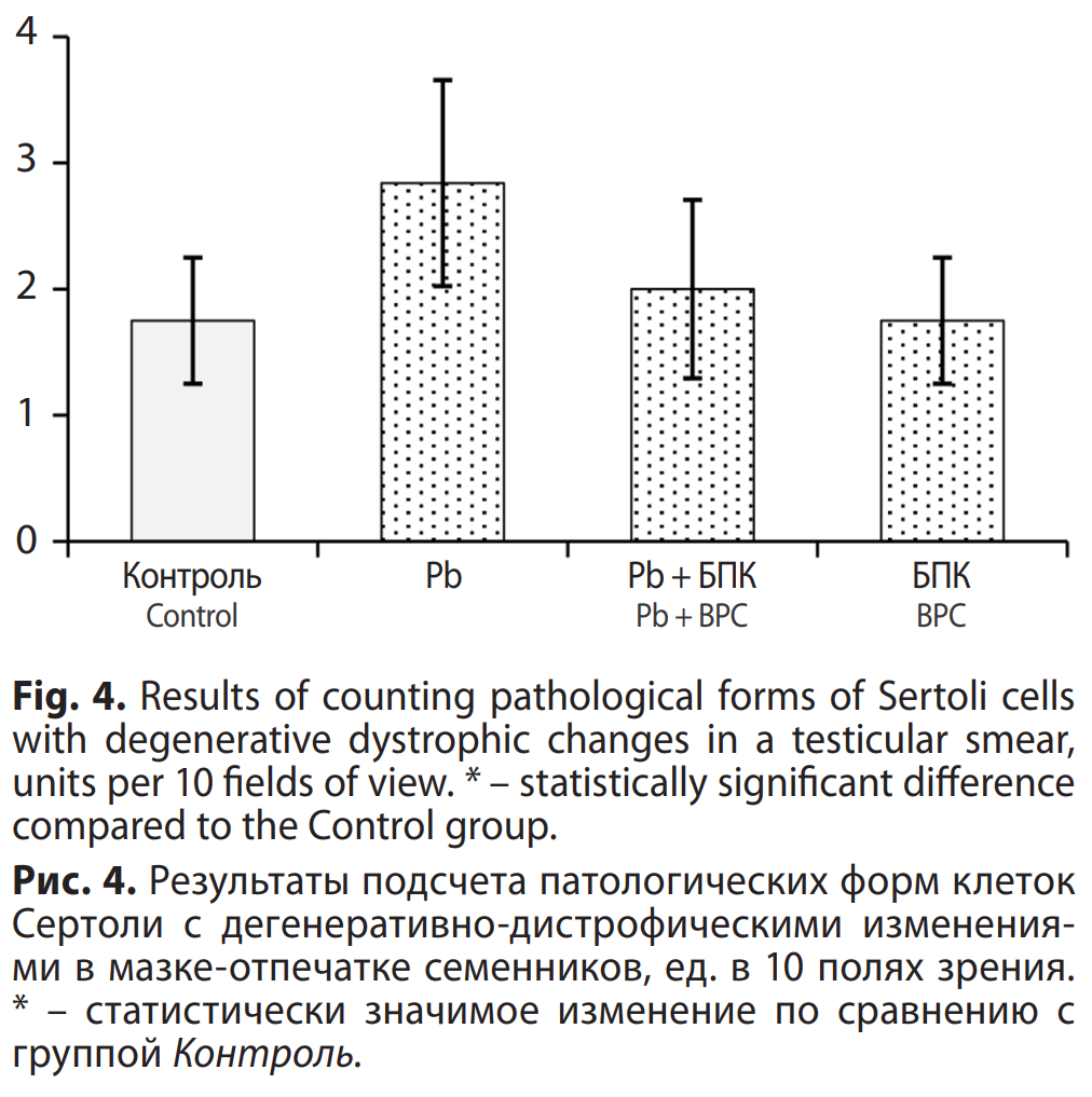
Analysis of testicular smears in the Pb group revealed a tendency to increase the number of degeneratively altered Sertoli cells. The Pb + BPC and BPC groups demonstrated values equivalent to those of the Control (Fig. 4).
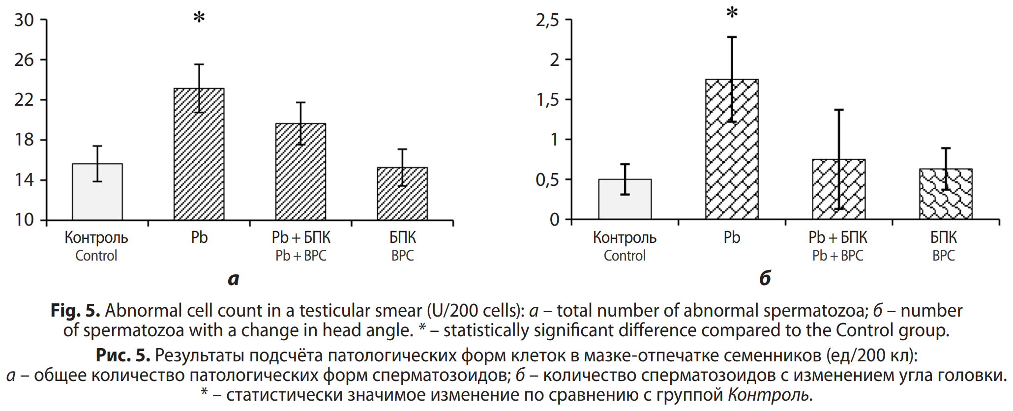
A significant increase in abnormal sperm forms, particularly cells with altered head angles, was observed in the Pb group, compared to the control. However, changes in the Pb + BPC group were less pronounced or did not differ at all from control values (Fig. 5, a, b).
Discussion
One of the key regulators of sex hormone synthesis is the membrane-bound enzyme 3bHSD from the oxidoreductase class [4,5]. Several studies demonstrate that exposure to inorganic lead compounds results in a reduction of the 3bHSD enzyme level in the testes [6–8]. Meanwhile, the increase in the content of the HSD3b1 isoform of the 3bHSD enzyme in blood serum observed in the current study could be the result of the destruction of cells involved in androgen synthesis due to toxic lead exposure.
Leydig cells are known to be responsible for the synthesis of male sex hormones, particularly testosterone [10, 11]. The highest number of Leydig cells is observed during puberty, but their number gradually decreases with age. Testosterone is one of the main regulators of spermatogenesis and sexual function in general [12, 13]. In this study, we did not determine sex hormone levels, which does not allow us to objectively assess the disruption of androgen synthesis. Nevertheless, the results of the histomorphometric analysis suggest a positive effect of BPC on the functional activity of spermatogenesis mechanisms in exposed male rats.
it should be noted that changes in spermatogenesis parameters are likely related not only to the direct effect of lead acetate on seminal cells, but also to disturbances in the hypothalamic-pituitary system, which is responsible for neuroendocrine regulation of the reproductive system. Lead is known to inhibit the synthesis of luteinizing hormone [14, 15] in the anterior pituitary gland, one of whose main functions is to stimulate testosterone synthesis. This hypothesis is also supported by the results of our previous studies, which demonstrated the pronounced toxic effect of lead acetate on the central nervous system of laboratory animals and the neuroprotective effect of BPC [16, 17]. The identified changes only provide indirect evidence of impaired neurohumoral regulation, and confirmation of this hypothesis requires determination of sex hormone levels. However, based on the parameters studied, the developed BPC demonstrated a protective effect against lead acetate exposure.
Limitations of the study. We did not assess sex hormone levels, as well as the content of the HSD3b2 isoform of the 3bHSD enzyme. To complete the study and substantiate the genotoxic risks of the factor under study, it is advisable to determine the expression of the HSD3B1 and HSD3B2 genes in the testes and adrenal glands. To determine the possible reasons of an increase in the content of the HSD3b1 enzyme in the blood, it is necessary to assess its level in tissues involved in the synthesis of androgens. We were limited to the use of a single test substance, route of administration, and exposure time.
Conclusion
According to the study results, male rats exposed to lead acetate showed a slight increase in the blood content of the enzyme 3b-hydroxysteroid dehydrogenase, which plays an important role in regulating sex hormone synthesis. The observed histomorphometric changes included a decrease in the lumen of the seminiferous tubules, an increase in the number of abnormal Sertoli cells and degenerated spermatozoa. As a whole, the experimental findings confirmed the protective effect of the bioprophylactic complex based on histomorphometric parameters. The findings revealed the protective potential of biological prevention strategies but highlight the importance of further profound studies of the mechanisms of lead toxicity on the male reproductive system and the effects of of the developed bioprophylactic complex (BPC).
¹ On the State of Sanitary-Epidemiological Well-Being of the Population in the Russian Federation in 2024: State Report. Moscow: Federal Service for Supervision in Healthcare Federal Service for Supervision on Consumer Rights Protection and Human Wellbeing, 2025. 424 p.
² GOST 33216–2014 Guidelines for Accommodation and Care of Animals. Species-Specific Provisions for Laboratory Rodents and Rabbits.
³ GOST R 50258–92 Complete Compound Feeds for Laboratory Animals. Specifications.
1. Sutunkova M.P., Minigalieva I.A., Gertan N.A., Petrunina E.M., Tsypushkina E.E., Nikogosyan K.M. Bioprophylaxis of the reprotoxic effect of sulfuric acid: an experimental study. Meditsina truda i ekologiya cheloveka. 2025; (2): 111–24. https://doi.org/10.24412/2411-3794-2025-10208 https://elibrary.ru/mgnaah (in Russian)
2. Privalova L.I., Klinova S.V., Minigalieva I.A., Ryabova Iu.V., Sutunkova M.P., Makeev O.G., et al. An experimental trial of bioprophylactic formula designed to minimize combined toxicity of both lead and cadmium. Gigiena i Sanitaria (Hygiene and Sanitation, Russian journal). 2020; 99(1): 85–9. https://elibrary.ru/yzqcos (in Russian)
3. Klinova S.V., Minigalieva I.A., Sutunkova M.P., Privalova L.I., Gerzen O.P., Riabova Yu.V., et al. Effects of lead and/or cadmium on the contractile function of the rat myocardium following subchronic exposure and its attenuation with a complex of bioprotectors. Zdorov’e naseleniya i sreda obitaniya – ZNiSO. 2021; 29(6): 25–33. https://elibrary.ru/lmooqz (in Russian)
4. Simard J., Ricketts M.L., Gingras S., Soucy P., Feltus F.A., Melner M.H. Molecular biology of the 3beta-hydroxysteroid dehydrogenase/delta5-delta4 isomerase gene family. Endocr. Rev. 2005; 26(4): 525–82. https://doi.org/10.1210/er.2002-0050
5. Simard J., Couet J., Durocher F., Labrie Y., Sanchez R., Breton N., et al. Structure and tissue-specific expression of a novel member of the rat 3 beta-hydroxysteroid dehydrogenase/delta 5-delta 4 isomerase (3 beta-HSD) family. The exclusive 3 beta-HSD gene expression in the skin. J. Biol. Chem. 1993; 268(26): 19659–68. https://doi.org/10.1016/S0021-9258(19)36567-6
6. Anjum M.R., Sainath S.B., Suneetha Y., Reddy P.S. Lead acetate induced reproductive and paternal mediated developmental toxicity in rats. Ecotoxicol. Environ. Saf. 2011; 74(4): 793–9. https://doi.org/10.1016/j.ecoenv.2010.10.044
7. Sainath S.B., Meena R., Supriya Ch., Reddy K.P., Reddy P.S. Protective role of Centella asiatica on lead-induced oxidative stress and suppressed reproductive health in male rats. Environ. Toxicol. Pharmacol. 2011; 32(2): 146–54. https://doi.org/10.1016/j.etap.2011.04.005
8. Khafaji S.S. Antioxidant, anti-inflammatory, and anti-reprotoxic effects of kaempferol and vitamin E on lead acetate-induced testicular toxicity in male rats. Open Vet. J. 2023; 13(12): 1683–95. https://doi.org/10.5455/OVJ.2023.v13.i12.17
9. Tomita T., Sato T., Saito K., Takakuwa E. Effects of lead and arsenic on the formation of 5β-H steroids. Toxicol. Lett. 1979; 3(5): 291–7. https://doi.org/10.1016/0378-4274(79)90006-7
10. Zirkin B.R., Papadopoulos V. Leydig cells: formation, function, and regulation. Biol. Reprod. 2018; 99(1): 101–11. https://doi.org/10.1093/biolre/ioy059
11. Bakhtyukov A.A., Shpakov A.O. The molecular mechanisms of steroidogenesis regulation in Leydig cells. Tsitologiya. 2016; 58(9): 666–78. https://elibrary.ru/wjljej (in Russian)
12. Grande G., Barrachina F., Soler-Ventura A., Jodar M., Mancini F., Marana R., et al. The role of testosterone in spermatogenesis: lessons from proteome profiling of human spermatozoa in testosterone deficiency. Front. Endocrinol. (Lausanne). 2022; 13: 852661. https://doi.org/10.3389/fendo.2022.852661
13. Smith L.B., Walker W.H. The regulation of spermatogenesis by androgens. Semin. Cell Dev. Biol. 2014; 30: 2–13. https://doi.org/10.1016/j.semcdb.2014.02.012
14. Al-Megrin W.A., Alomar S., Alkhuriji A.F., Metwally D.M., Mohamed S.K., Kassab R.B., et al. Luteolin protects against testicular injury induced by lead acetate by activating the Nrf2/HO-1 pathway. IUBMB Life. 2020; 72(8): 1787–98. https://doi.org/10.1002/iub.2311
15. Khalaf M.A.M., Younis R.H.A., El-Fakahany H. Effect of low-level environmental lead exposure on the onset of male puberty. Int. J. Toxicol. 2019; 38(3): 209–14. https://doi.org/10.1177/1091581819848411
16. Minigalieva I.A., Nikogosyan K.M., Sutunkova M.P., Kungurtseva A.K., Petrunina E.M., Bateneva V.A. Experimental evaluation of a bioprophylactic complex aimed at increasing resistance to neurotoxic effects of lead oxide nanoparticles. Toksikologicheskii vestnik. 2025; 33(2): 86–92. https://doi.org/10.47470/0869-7922-2025-33-2-86-92 https://elibrary.ru/bgxhph (in Russian)
17. Ryabova Yu.V., Kungurtseva A.K., Petrunina E.M., Nikogosyan K.M., Klinova S.V., Minigalieva I.A., et al. Changes in health effects of lead exposure caused by exercise and the impact of biological prophylaxis on rats’ central nervous system. Meditsina truda i ekologiya cheloveka. 2024; (2): 191–210. https://doi.org/10.24412/2411-3794-2024-10213 https://elibrary.ru/
Researcher at the Department of Toxicology and Bioprophylaxis, Yekaterinburg Medical Research Center for Prophylaxis and Health Protection of Industrial Workers of Rospotrebnadzor, Yekaterinburg, 620014, Russian Federation
e-mail: nikoghosyankm@ymrc.ru
Doctor of Medical Sciences, Director of the Yekaterinburg Medical Research Center for Prophylaxis and Health Protection of Industrial Workers of Rospotrebnadzor, Yekaterinburg, 620014, Russian Federation; Head of the Department of Occupational Hygiene and Medicine, Ural State Medical University of the Ministry of Health of the Russian Federation, Yekaterinburg, 620028, Russian Federation
e-mail: sutunkova@ymrc.ru
Doctor of Biological Sciences, Head of the Department of Toxicology and Bioprophylaxis, Yekaterinburg Medical Research Center for Prophylaxis and Health Protection of Industrial Workers of Rospotrebnadzor, Yekaterinburg, 620014, Russian Federation
e-mail: ilzira@ymrc.ru
Junior Researcher at the Department of Toxicology and Bioprophylaxis, Yekaterinburg Medical Research Center for Prophylaxis and Health Protection of Industrial Workers of Rospotrebnadzor, Yekaterinburg, 620014, Russian Federation
e-mail: gertanna@ymrc.ru
Junior Researcher at the Department of Toxicology and Bioprophylaxis, Yekaterinburg Medical Research Center for Prophylaxis and Health Protection of Industrial Workers of Rospotrebnadzor, Yekaterinburg, 620014, Russian Federation
e-mail: ekaterina_b89@list.ru
Candidate of Medical Sciences, Head of the Department of Laboratory and Diagnostic Technologies, Yekaterinburg Medical Research Center for Prophylaxis and Health Protection of Industrial Workers of Rospotrebnadzor, Yekaterinburg, 620014, Russian Federation
e-mail: sahautdinova@ymrc.ru
Nikogosyan K.M., Sutunkova M.P., Minigalieva I.A., Gertan N.A., Petrunina E.M., Sakhautdinova R.R. Changes in some parameters of the reproductive system of male rats following lead exposure and the effect of bioprophylaxis. Toxicological Review. 2026;34(2):124-131. https://doi.org/10.47470/0869-7922-2026-34-2-124-131. EDN: xsvivv
 Email this article (Login required)
Email the author (Login required)
Email this article (Login required)
Email the author (Login required) 8 build 2, Bagrationovsky proezd, 121087 Moscow, Russian Federation
Branch of «Russian Register of Potentially Hazardous Chemical and Biological Substances» FBES F.F. Erisman Federal Scientific Center of Hygiene of Rospotrebnadzor
tel.: +7 (499) 145 60 23 (reception)
e-mail: director@rosreg.info
фыв